Looking Good How To Read A Breast Ultrasound Report
LOL The findings were.
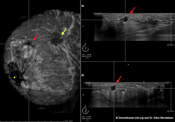
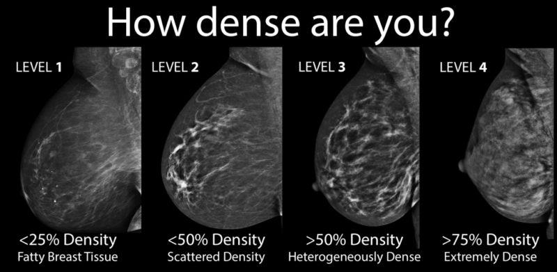
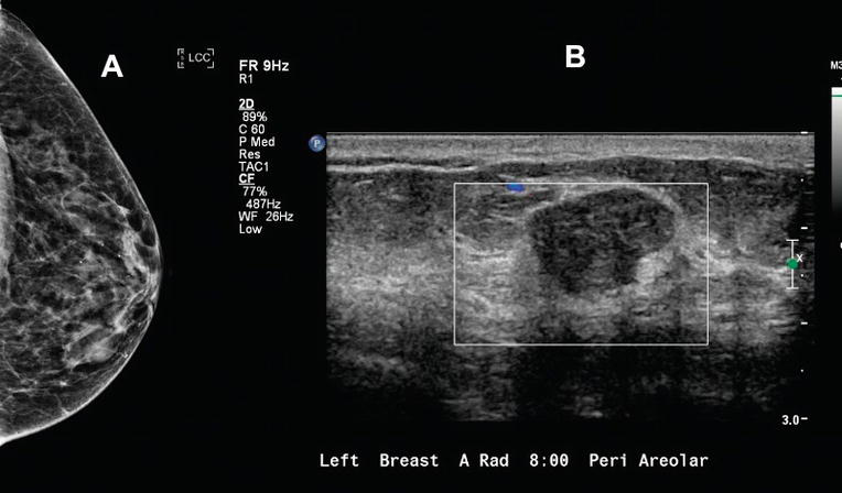
How to read a breast ultrasound report. It is used to help diagnose breast lumps or other abnormalities found during a physical exam or on a mammogram or breast MRI. This conductive gel helps the ultrasound waves travel through your skin. The X-ray image below shows a suspect breast mass of about 1 cm in diameter.
Each patients cancer. First the sonographer or doctor will ask you to undress from the waist up and lie on your back on an ultrasound table. The anterior margin of the tear is adjacent to.
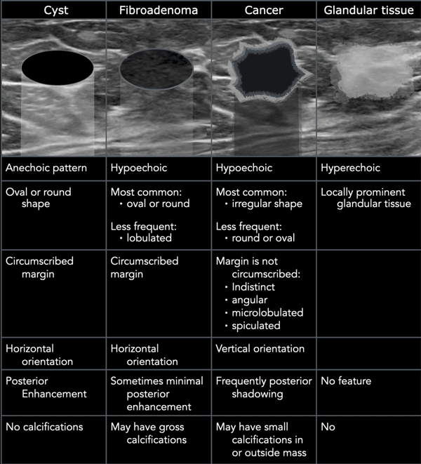
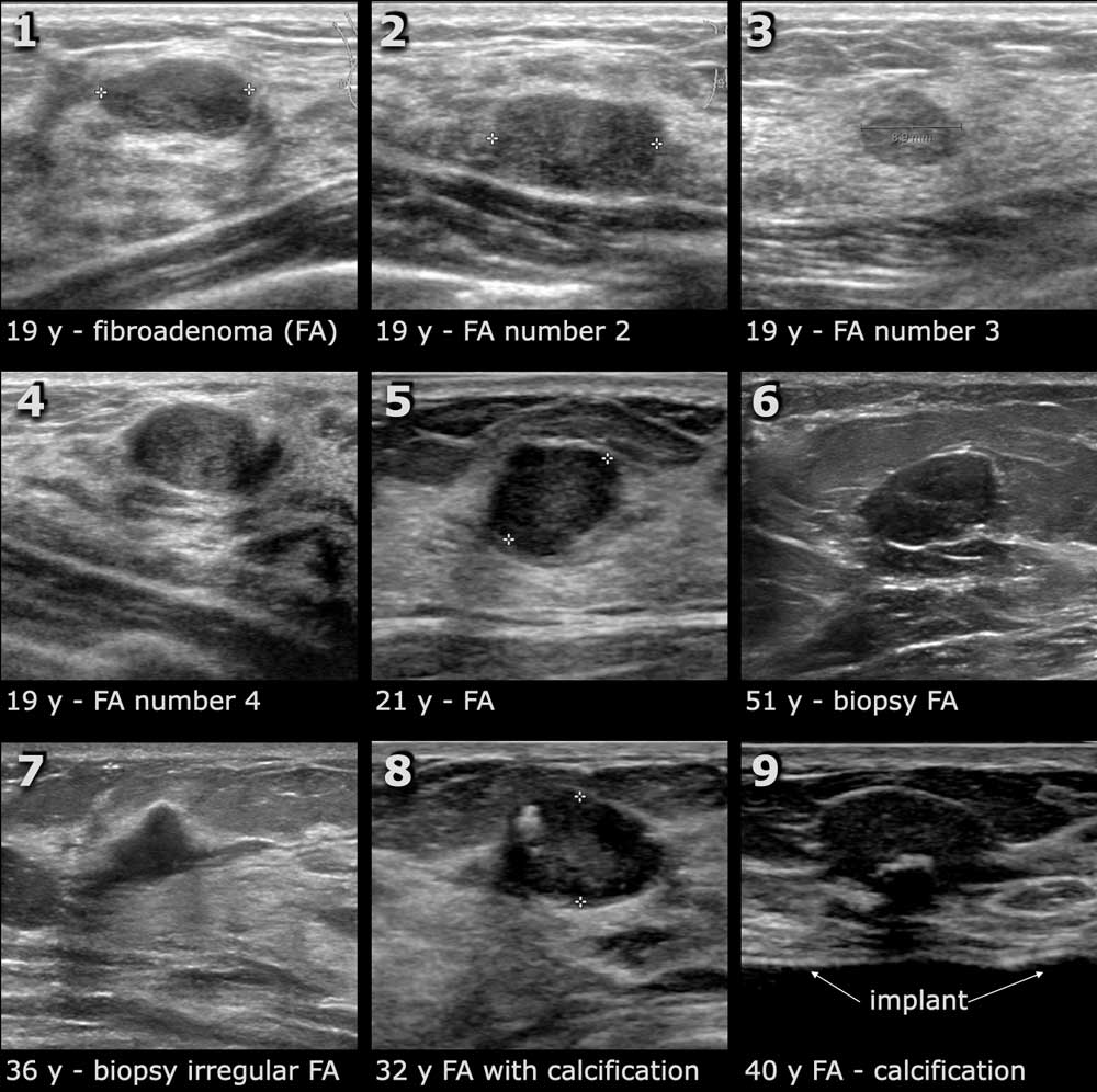
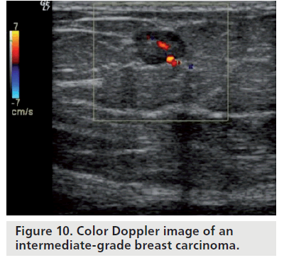
Its most important use however is to determine anomalies andgenetic disorders in the foetus early on in pregnancy that gives time for the doctors and the expectant mother to think about corrective measures even while the baby is in the womb. Breast FNA or Core Biopsy. The mass appears to be hypoechoic with ill-defined spiculated and microlobulated margins.
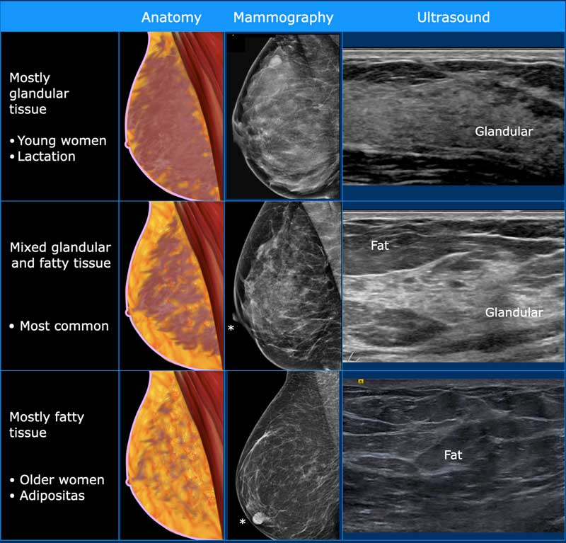
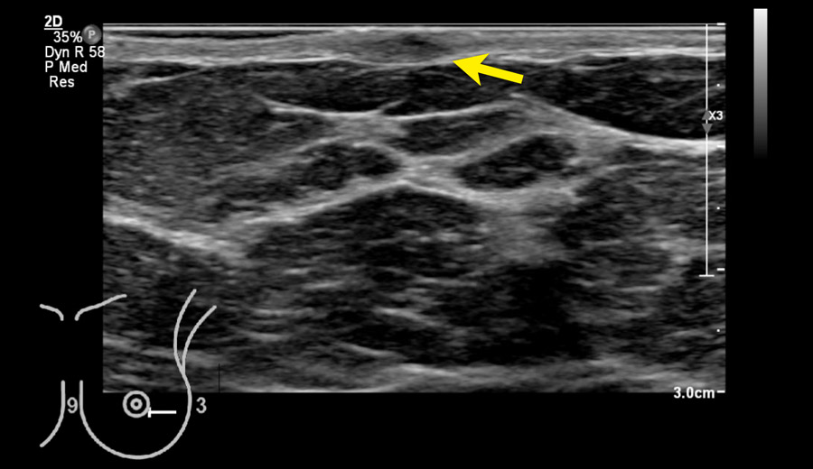
Here we see a normal ultrasound image of the breast. It is the most common cause of cancer death in women In 2005 alone 519 000 deaths were recorded due to breast cancer This means that one in every 100 deaths worldwide and almost one in every 15 cancer deaths were due to breast cancer. Figure 5-1 Normal breast ultrasound images.
The sound waves from the ultrasound travel right through it with very little bounce-back. Ultrasound imaging of the breast uses sound waves to produce pictures of the internal structures of the breast. Pathologists evaluate the tissue within a breast core biopsy or surgical specimen to diagnose the type of cancer as well as evaluate the presence of various prognostic factors.
During my mammo a tumor was foundI was asked to wait same day for a ultrasoundThe ultrasound was just to make sure what it could beCyst tumor or maybe nothingI had the ultrasound that same dayDoctor read the Xray and said it was a tumor and he didnt think it was anything BUT I must have a biopsyAfter the biopsy is scheduled you usually know in a couple days. On your report the radiologist may describe enhancement or may describe an enhancing mass. To read an ultrasound picture look for white spots on the image to see solid tissues like bones and dark spots on the image to see fluid-filled tissues like the amniotic fluid in the uterus.
